Ultrasound examinations in infancy
Brain ultrasound examination of infants
With this method, the brain tissue can be examined through the large well. During the examination, the spaciousness of the cerebral ventricles is checked, and extensive pathological deviations in the brain tissue, as well as some developmental disorders, can also be detected by ultrasound. Vascular abnormalities, cysts, and brain haemorrhages that do not cause symptoms may be revealed. In the case of difficult births, developmental and movement disorders in infancy, an ultrasound examination of the brain is also of particular importance. In the case of clinical symptoms, or if the possibility of brain damage has arisen during childbirth, in the early new-born age, it is definitely justified to carry out the examination.
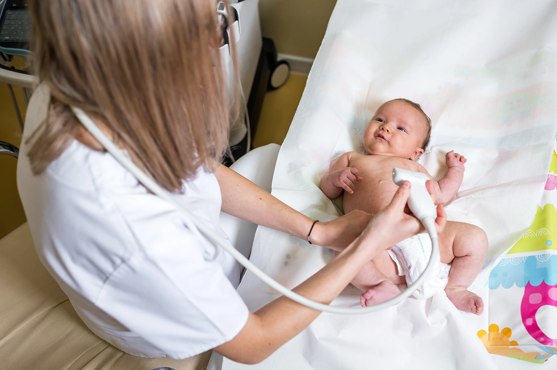
Abdominal ultrasound examination of infants
During the examination, we look at all the abdominal organs, especially the kidneys, adrenal glands, urinary tract, liver, and bile ducts. In this way, possible congenital abnormalities can be easily detected.
Abdominal examination is most important in detecting abnormalities of kidney development in infants without complaints. The ultrasound examination is a fast, effective and safe method to examine the size, position and structure of the kidneys, whether both kidneys are present at all, and whether there is an expansion of the cavity system. In this way, possible pathological deviations can be recognized in time, and if necessary, treatment can be started immediately. The test is also very important for babies who already had visible dilatation of the cavity system in the kidney during the fetal-age, or whose family has a history of such a hereditary disease.
In the case of boys, the undescended testicles can be found and their position determined with the ultrasound examination, and the abdominal and pelvic lymph nodes can also be filtered exceptionally well.
In the case of jaundice, it is essential to examine the bile ducts, and to rule out gastric stricture in babies with vomiting and abdominal pain.
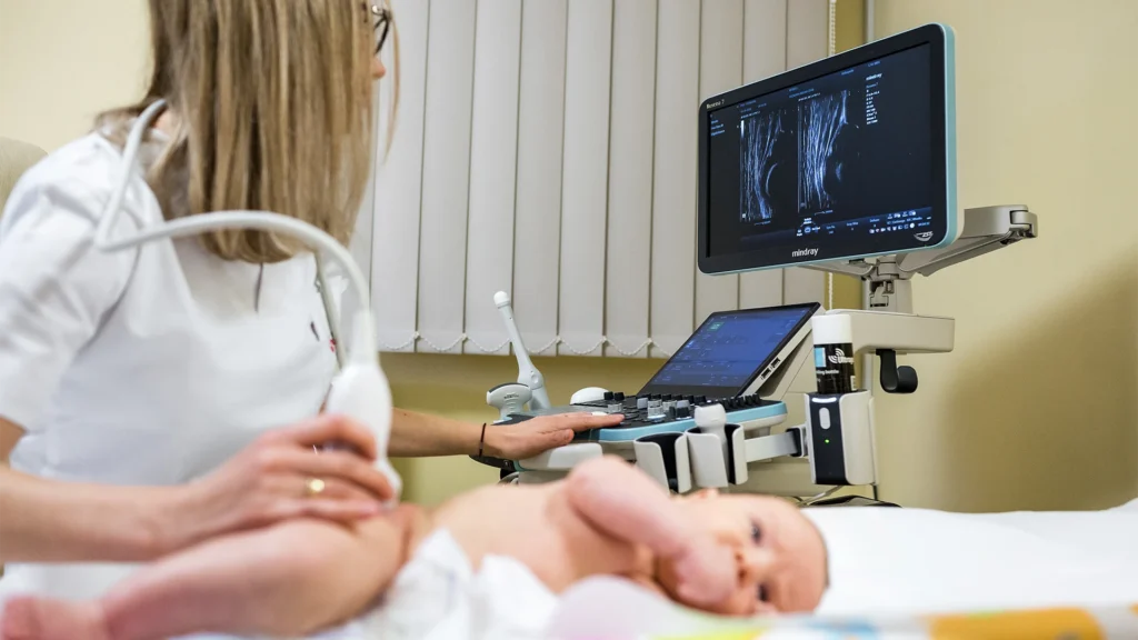
Hip screening of babies with ultrasound examination
An ultrasound examination of the hip gives a more accurate and precise diagnosis than a clinical examination, and does not burden the new-born with X-rays.
The ideal age for hip ultrasound is 6-8 weeks. In most cases, the deviation detected at this age can be corrected with a simple harness or a diaper with a spread insert.
This test is especially recommended for babies with a family history of hip sprains or breech births during pregnancy.
If we recognize possible deviations in time (e.g. an underdeveloped hip joint or a hip joint prone to hip sprains), we can start treatment early, which in most cases consists of the use of diapers with splayed inserts or different stirrups, which are not painful for the baby. In these cases, we also carry out a control examination, and if during this we do not notice sufficient improvement – which fortunately rarely happens – we recommend a paediatric orthopaedic examination.
Screening for heart abnormalities in babies using ultrasound
Congenital heart defects are among the most frequently detected developmental defects. The spectrum of the disease is very broad, ranging from minimal structural deviations to complex cases requiring surgical intervention.
During the ultrasound examination of the heart, we get information about the child’s heart, the functioning of the heart valves, their structure, size, congenital or acquired abnormalities, performance, and possible wall thickening. The fluid accumulated in the pericardium can be represented. The normal or abnormal flow of blood between the heart chambers can be displayed in color.
With ultrasound examination, the progression or regression of possible developmental disorders can be easily followed, so it is possible to determine the time of diseases requiring intervention in time.